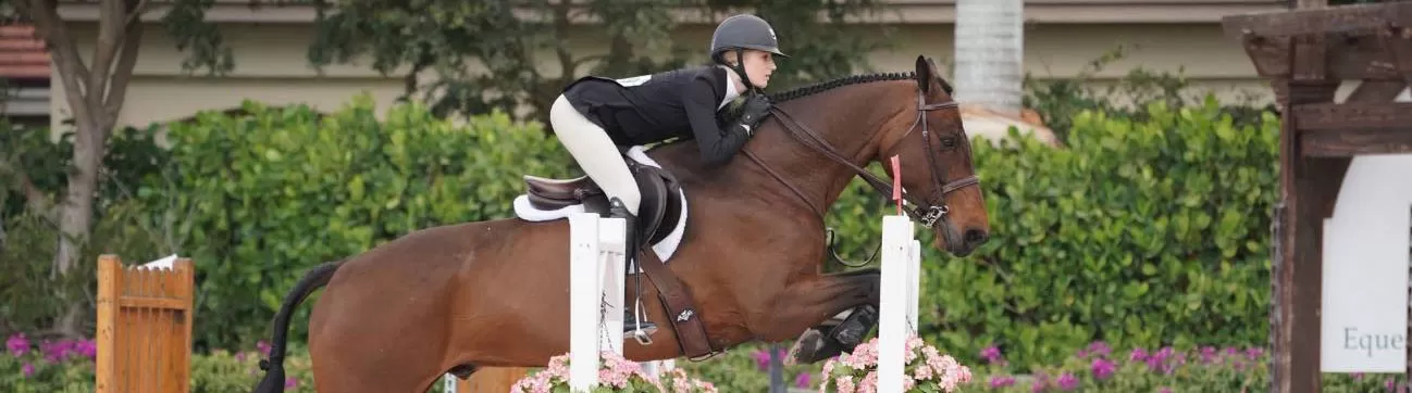
 We are happy that your horse is going home and getting back to a familiar environment! Now that surgery is completed, your horse will require special attention during the recovery period. Follow-up care at home is an essential part of successful joint therapy and will contribute significantly to the eventual return of your horse to performance soundness.
[ezcol_1half]
 [/ezcol_1half][ezcol_1half_end]
 [/ezcol_1half_end]
STALL REST
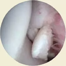
Since arthroscopic incisions are relatively small, movement usually has a minimal effect on incisional healing of the skin. However, stall rest is required in post-operative arthroscopic patients to allow joint inflammation to subside and cartilage/bone healing to commence. Generally, horses are stall rested for 7 days after surgery to enable the blood clot which has formed in the cartilage/bone defect(s) to organize and to allow granulation tissue to begin forming. Handwalking after this period helps the clot to further organize and create a smooth gliding surface within the joint. Rehabilitation regimens differ for horses of different ages, type of work, breed, size, type of lesion(s), and/or location of lesion(s). Following are typical recovery schedules for post-operative arthroscopic patients based on type and location of the lesion(s):
CARPUS:
- Chip Fractures – Stall rest only for 7 days. After 7 days passive joint flexion (10 minutes twice daily) and hand walking (10 minutes twice daily) can begin.
- If fragmentation within the joint is simple and fresh, training may begin at 6 weeks.
- Horses with evidence of mild-to-moderate cartilage degeneration within the joint require a minimum of 3 months stall rest with handwalking before training may resume.
- Horses with severe cartilage degeneration should have 4 to 6 weeks rest.
- Slab Fractures – Stall rest only for 7 days. After 7 days an increasing regimen of handwalking for the following 8 weeks. This is followed by a minimum of 3 months of turnout prior to resuming work.
FETLOCK:
- Chip Fractures – Stall rest only for 7 days. After 7 days passive joint flexion (10 minutes twice daily) and hand walking (10 minutes twice daily) can begin.
- If fragmentation within the joint is simple and fresh, training may begin at 6 weeks.
- Depending on the extent of involvement, convalescent time may be increased to up to 6 months
- Chronic Proliferative (Villonodular) Synovitis – Stall rest only for 7 days. After 7 days passive joint flexion (10 minutes twice daily) and hand walking (10 minutes twice daily) can begin. This should be continued for at least 90 days before work resumes.
- OCD Lesions of the Midsaggital Ridge – Stall rest only for 7 days. After 7 days passive joint flexion (10 minutes twice daily) and hand walking (10 minutes twice daily) can begin.
- If fragmentation within the joint is simple and fresh, training may begin at 6 weeks.
- Depending on the extent of involvement, convalescent time may be increased to up to 6 months.
- OCD Lesions of Proximal Palmar (Plantar) First Phalanx – Stall rest only for 7 days. After 7 days passive joint flexion (10 minutes twice daily) and hand walking (10 minutes twice daily) can begin. This should be continued for 60 to 90 days before work resumes.
- Apical Fragments of the Proximal Sesamoid Bones – Stall rest only for 7 days. After 7 days passive joint flexion (10 minutes twice daily) and hand walking (10 minutes twice daily) can begin. Depending on the fragment size and appearance of the joint at the time of surgery, stall rest with handwalking is continued for 1 to 6 months.
STIFLE:
- OCD Lesions of the Lateral and Medial Trochlear Ridges and the Patella – Stall rest only for 7 days. After 7 days hand walking (10 minutes twice daily) can begin. This should be continued for 90 to 120 days before work resumes.
- Fragmentation of the Distal Patella – Stall rest only for 7 days. After 7 days passive joint flexion (10 minutes twice daily) and hand walking (10 minutes twice daily) can begin. This should be continued for 6 months before work resumes.
- Subchondral Cystic Lesion of the Medial Femoral Condyle – Stall rest only for 14 days. After 14 days passive joint flexion (10 minutes twice daily) and hand walking (10 minutes twice daily) can begin and is continued for 60 days. Turnout for 120 days is recommended before work resumes.
SHOULDER:
- OCD Lesions of the Scapulohumeral Joint – Stall rest only for 10-14 days. After 10-14 days, handwalking (5 minutes per day) can begin. The period of hand walking is increased by increments of 5 minutes weekly until the horse has walked for 30 minutes each day for one week. After this time, the horse is turned out for 4 to 12 months (depending on the age of the animal and severity of the lesion(s) before work resumes.)
TARSUS:
- OCD Lesions of the Distal Intermediate Ridge of the Tibia – Stall rest only for 7 days. After 7 days passive joint flexion (10 minutes twice daily) and hand walking (10 minutes twice daily) can begin and is continued for 4 weeks. After this, small paddock turnout or controlled light exercise is allowed for another 4 weeks. Light training may resume in 9 weeks post-operatively.
- OCD Lesions of the Trochlear Ridges – Stall rest only for 7 days. After 7 days passive joint flexion (10 minutes twice daily) and hand walking (10 minutes twice daily) can begin and is continued for 4 weeks. After this, small paddock turnout or controlled light exercise is allowed for another 4 weeks. Light training may resume in 9 weeks post-operatively.
- OCD Lesions of the Medial Malleolus – Stall rest only for 7 days. After 7 days passive joint flexion (10 minutes twice daily) and hand walking (10 minutes twice daily) can begin and is continued for 4 weeks. After this, small paddock turnout or controlled light exercise is allowed for another 4 weeks. Light training may resume in 9 weeks post-operatively.
- Fragmentation of the Lateral Malleolus – Stall rest only for 7 days. After 7 days passive joint flexion (10 minutes twice daily) and hand walking (10 minutes twice daily) can begin and is continued for 4 weeks. After this, small paddock turnout or controlled light exercise is allowed for another 4 weeks. Light training may resume in 9 weeks post-operatively.
FURTHER TREATMENT
Most horses undergoing arthroscopic surgery of one or more joints already have associated inflammation in those areas. Joint inflammation occurs when irritated or inflamed synovium releases immune cells into the synovial fluid. Immune cells may subsequently release enzymes which can degrade normal cartilage. This process contributes to “degenerative joint disease”, and can result in chronic unsoundness in horses. Therefore, our primary goal is to eliminate as many sources of inflammation as possible. Although surgery has already helped to do this, the surgical procedure itself may result in an inflammatory response by your horse. For this reason, horses undergoing arthroscopic surgery are placed on a 14-day post-operative regimen of phenylbutazone (an anti-inflammatory agent).
Joint health may also be improved by intra-articular and/or systemic treatment with medications designed to normalize the synovial environment. Such medications include Hyaluartin V (intra-articular), Legend (intravenous), Cosequin (oral), and Adequan (intramuscular). These medications may help to improve the health of the synovial membrane and therefore the joint. Hyaluratin V was injected into your horse’s joint(s) at the time of surgery, and Legend was administered the morning of discharge. Although further treatment may not be necessary to keep your horse sound, we encourage an Adequan and/or Cosequin treatment regimen to begin after your horse goes home. Please ask your attending veterinarian about utilizing these products for your horse.
PROGNOSIS
In most cases, prognosis for future performance is closely related to the amount of degenerative change that has already occurred within the joint(s) involved. Typically, the longer a lesion has been present the more degenerative change has occurred. Consequently, fresh injuries often have a better prognosis than chronic injuries. Since cartilage has little or no regenerating capacity in adult horses, degenerative cartilage will likely remain abnormal for the remainder of the animal’s life. An exception might be evident in very young horses and foals. In most cases, however, degenerative change within the joint cannot be reversed. Proper treatment, therefore, is designed to prevent further joint degeneration and inflammation. Surgery coupled with aggressive peri-operative medication can help us to achieve this goal. Although many aspects of degenerative joint disease (DJD) are evident on pre-operative radiographs, lesions solely involving the cartilage are radiographically invisible and are much more accurately assessed by visualization through an arthroscope. Therefore, we typically wait to give a final prognosis until after the arthroscopic procedure. It follows that the less degenerative changes that are apparent within a joint at the time of surgery, the better will be the prognosis for return to future performance.
Following is a chart that was produced from the results of arthroscopic surgery on hundreds of horses. It may help to give you an idea of percentage of success regarding specific arthroscopic lesions.
| AREA OF LESION | TYPE OF LESION | BREED(S) OF HORSE | SUCCESS* RATE |
|
|
|
|
|
| Distal Radial Carpal Bone (Carpus) | chip fracture | QH | 70.6% |
| TB | 55.4% | ||
| Distal Intermediate Carpal Bone (Carpus) | chip fracture | QH | 80% |
| TB | 99% | ||
| Proximal 3rd Carpal Bone (Carpus) | chip fracture | QH | 29.4% |
| TB | 58.8% | ||
| Distal Radius (Carpus) | chip fracture | QH | 80% |
| TB | 74% | ||
| Proximal Intermediate Carpal Bone (Carpus) | chip fracture | QH | 89.7% |
| TB | 61.5% | ||
| Proximal Radial Carpal Bone (Carpus) | chip fracture | QH | 99% |
| TB | 75% | ||
| Medial Palmar Intercarpal Ligament (Carpus) | partial tear | ALL | 50% |
| 3rd Carpal Bone (Carpus) | slab fracture | ALL | 50% |
| Proximal First Phalanx (Fetlock) | chip fracture | QH, TB | 86.5% |
| Proximal Fetlock Joint | proliferative synovitis | ALL | guarded |
| AREA OF LESION | TYPE OF LESION | BREED(S) OF HORSE | SUCCESS* RATE |
|
|
|
|
|
| Proximal Midsaggital Ridge (Fetlock) | OCD lesion | ALL | depends on joint appearance |
| Proximal Palmar (Plantar) First Phalanx | OCD lesion | ALL | depends on joint appearance |
| Proximal Sesamoid Bone | apical fracture | ALL | guarded |
| Lateral and Medial Trochlear Ridge, Patella (Femoropatellar Joint) | OCD lesion | ALL | 80% |
| Distal Patella | fragmentation | ALL | 80% |
| Medial Femoral Condyle | subchondral cyst | ALL | 72% |
| Distal Intermediate Ridge of the Tibia | OCD lesion | ALL | 85% |
| Medial Malleolus of the Tibia | OCD lesion | ALL | 85% |
| Lateral/Medial Trochlear Ridges of the Tibia | OCD lesion | ALL | 85% |
| Scapulohumeral Joint | OCD lesion | ALL | 81.8% |
*This represents the percentage of horses that returned to a level of performance equal to or better than the preinjury level.